 The pipeline for EM + Atlas based brain tissue segmentation.
Abstract
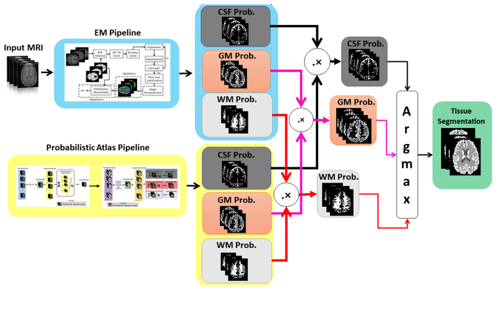
Brain tissue segmentation is one of the primary tools in analysing brain tissues. In previous labs, Expectation Maximization (EM) and Atlas construction strategies, which are widely-used approaches for brain tissue segmentation, were implemented from scratch. This course-work is a continuation of our registration lab where we built probabilistic atlas for brain tissue segmentation. Here, We are combining EM with Atlas for segmenting Cerebrospinal fluid (CSF) with label 1, White Matter (WM) with label 2, and Gray Matter (GM) with label 3. In this report, we propose a solution of 5 complexity levels, namely:
- Computing the probabilistic atlas of the available dataset.
- Finding the intensity distribution of the training images for each region.
- Implementing the Bayesian framework by joining the upper two steps.
- Using step 3, initialise EM and compute per-region voxel probabilities.
- The joint framework, Bayesian EM: combining EM and Bayesian framework.
Type
Publication
MAIA Medical Image Segmentation and Applications Course